Unveiling The Secrets Of Astigmatism: A Journey Of Clarity
Astigmatism Pictures
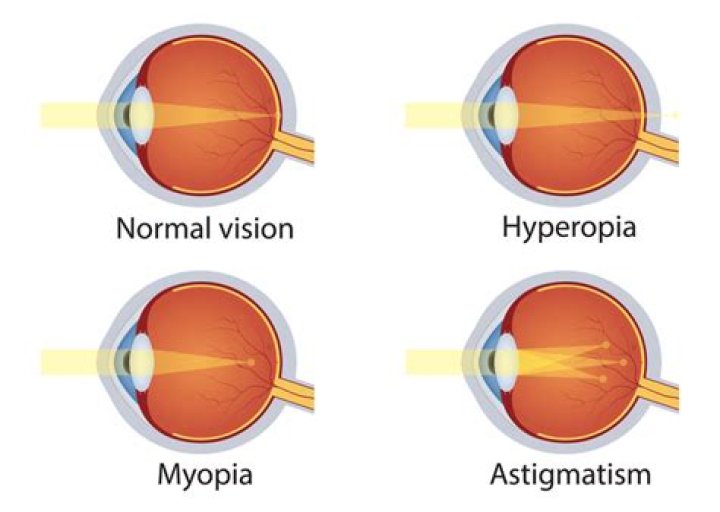
Astigmatism is a common vision condition that occurs when the cornea, the clear outer layer of the eye, is not perfectly round. This causes light to be focused in two different places on the retina, resulting in blurred vision.
Astigmatism pictures are images that are used to diagnose and monitor astigmatism. These pictures can be taken with a variety of devices, including autorefractors, keratometers, and topographers.
Autorefractors are used to measure the refractive error of the eye, which is the amount of light that is bent when it enters the eye. Keratometers are used to measure the curvature of the cornea. Topographers are used to create a map of the corneal surface.
Astigmatism pictures are an important tool for diagnosing and monitoring astigmatism. They can help doctors to determine the severity of the condition and to prescribe the appropriate treatment.
Astigmatism Pictures
Astigmatism pictures are an important tool for diagnosing and monitoring astigmatism, a common vision condition that occurs when the cornea, the clear outer layer of the eye, is not perfectly round. Astigmatism pictures can be taken with a variety of devices, including autorefractors, keratometers, and topographers.
- Diagnosis: Astigmatism pictures can help doctors to diagnose astigmatism and determine the severity of the condition.
- Monitoring: Astigmatism pictures can be used to monitor the progression of astigmatism over time.
- Treatment: Astigmatism pictures can help doctors to prescribe the appropriate treatment for astigmatism, such as glasses or contact lenses.
- Autorefraction: Autorefractors are used to take astigmatism pictures that measure the refractive error of the eye.
- Keratometry: Keratometers are used to take astigmatism pictures that measure the curvature of the cornea.
- Topography: Topographers are used to take astigmatism pictures that create a map of the corneal surface.
- Importance: Astigmatism pictures are an important tool for diagnosing and monitoring astigmatism because they can provide doctors with valuable information about the condition.
- Benefits: Astigmatism pictures can help doctors to provide patients with the best possible care.
- Availability: Astigmatism pictures are widely available and can be taken at most optometrists' offices.
Astigmatism pictures are an essential tool for diagnosing and monitoring astigmatism. They can help doctors to provide patients with the best possible care.
Diagnosis
Astigmatism pictures are an important tool for diagnosing astigmatism because they can provide doctors with a clear view of the cornea, the clear outer layer of the eye. This allows doctors to see if the cornea is irregularly shaped, which is the cause of astigmatism. Astigmatism pictures can also help doctors to determine the severity of the condition by measuring the amount of corneal irregularity.
- Corneal Topography: Corneal topography is a type of astigmatism picture that creates a map of the corneal surface. This map can show doctors the shape of the cornea and identify any areas of irregularity.
- Autorefraction: Autorefraction is another type of astigmatism picture that measures the refractive error of the eye. This information can help doctors to determine the severity of astigmatism and prescribe the appropriate corrective lenses.
- Keratometry: Keratometry is a type of astigmatism picture that measures the curvature of the cornea. This information can help doctors to determine the severity of astigmatism and prescribe the appropriate contact lenses.
- Slit-Lamp Examination: A slit-lamp examination is a type of eye exam that uses a bright light and a magnifying lens to examine the eye. This exam can help doctors to diagnose astigmatism and other eye conditions.
Astigmatism pictures are an essential tool for diagnosing and managing astigmatism. They can help doctors to provide patients with the best possible care.
Monitoring
Astigmatism pictures are an important tool for monitoring the progression of astigmatism over time. Astigmatism is a common vision condition that occurs when the cornea, the clear outer layer of the eye, is not perfectly round. This causes light to be focused in two different places on the retina, resulting in blurred vision.
Astigmatism pictures can be used to track changes in the shape of the cornea over time. This information can help doctors to determine whether astigmatism is getting worse or better. Astigmatism pictures can also be used to monitor the effectiveness of treatment for astigmatism.
For example, if a patient is wearing glasses or contact lenses to correct astigmatism, astigmatism pictures can be used to see if the lenses are working properly. Astigmatism pictures can also be used to monitor the progression of astigmatism in children. Astigmatism is a common condition in children, and it is important to monitor its progression to ensure that it does not affect their vision development.
Astigmatism pictures are an essential tool for monitoring the progression of astigmatism over time. They can help doctors to provide patients with the best possible care.
Treatment
Astigmatism pictures are an essential tool for diagnosing and managing astigmatism. They can help doctors to determine the severity of the condition and to prescribe the appropriate treatment.
- Glasses: Glasses are the most common treatment for astigmatism. They work by correcting the refractive error of the eye, which is the amount of light that is bent when it enters the eye. Glasses can be made with a variety of lenses, including spherical lenses, cylindrical lenses, and toric lenses.
- Contact lenses: Contact lenses are another option for correcting astigmatism. They work by resting directly on the surface of the eye. Contact lenses can be made with a variety of materials, including soft contact lenses, hard contact lenses, and gas permeable contact lenses.
- Surgery: In some cases, surgery may be necessary to correct astigmatism. Surgery can be used to reshape the cornea and improve vision.
Astigmatism pictures are an essential tool for helping doctors to prescribe the appropriate treatment for astigmatism. By providing a clear view of the cornea, astigmatism pictures can help doctors to determine the severity of the condition and to recommend the best course of treatment.
Autorefraction
Autorefraction is a method of measuring the refractive error of the eye using an autorefractor, a machine that shines a light into the eye and measures how the light is bent. Astigmatism is a common vision condition that occurs when the cornea, the clear outer layer of the eye, is not perfectly round. This causes light to be focused in two different places on the retina, resulting in blurred vision.
Astigmatism pictures are images of the eye that are taken with an autorefractor. These pictures can show the shape of the cornea and the amount of astigmatism. Astigmatism pictures are an important tool for diagnosing and managing astigmatism. They can help doctors to determine the severity of the condition and to prescribe the appropriate treatment.
Autorefraction is an important component of astigmatism pictures because it provides information about the refractive error of the eye. This information is used to create a prescription for glasses or contact lenses that will correct the astigmatism and improve vision.
Astigmatism pictures are an essential tool for diagnosing and managing astigmatism. They provide doctors with valuable information about the condition and help them to provide patients with the best possible care.
Keratometry
Keratometry is an important component of astigmatism pictures. It provides information about the curvature of the cornea, which is essential for diagnosing and managing astigmatism. Astigmatism is a common vision condition that occurs when the cornea is not perfectly round. This causes light to be focused in two different places on the retina, resulting in blurred vision.
Keratometers measure the curvature of the cornea by shining a light into the eye and measuring how the light is reflected. This information is used to create a map of the corneal surface. The map can show the shape of the cornea and identify any areas of irregularity. This information is essential for diagnosing and managing astigmatism.
Keratometry is a quick and painless procedure that can be performed in a doctor's office. It is an important part of a comprehensive eye exam and can help to ensure that patients receive the best possible care for their astigmatism.
Topography
Corneal topography is an important component of astigmatism pictures because it provides detailed information about the shape of the cornea. This information is essential for diagnosing and managing astigmatism, a common vision condition that occurs when the cornea is not perfectly round. Astigmatism causes light to be focused in two different places on the retina, resulting in blurred vision.
Topographers use a variety of techniques to create corneal topography maps. One common technique is to project a series of rings of light onto the cornea and measure how the rings are reflected. This information is used to create a map of the corneal surface, which can show the shape of the cornea and identify any areas of irregularity.
Corneal topography maps are essential for diagnosing and managing astigmatism. They can help doctors to determine the severity of the condition and to prescribe the appropriate treatment. Corneal topography maps can also be used to monitor the progression of astigmatism over time.
Astigmatism pictures are an important tool for diagnosing and managing astigmatism. Corneal topography is an essential component of astigmatism pictures because it provides detailed information about the shape of the cornea. This information is essential for diagnosing and managing astigmatism and ensuring that patients receive the best possible care.
Importance
Astigmatism pictures are essential for diagnosing and managing astigmatism, a common vision condition that occurs when the cornea, the clear outer layer of the eye, is not perfectly round. This causes light to be focused in two different places on the retina, resulting in blurred vision.
- Diagnosis: Astigmatism pictures can help doctors to diagnose astigmatism and determine the severity of the condition.
- Monitoring: Astigmatism pictures can be used to monitor the progression of astigmatism over time.
- Treatment: Astigmatism pictures can help doctors to prescribe the appropriate treatment for astigmatism, such as glasses or contact lenses.
Astigmatism pictures are an important tool for ensuring that patients receive the best possible care for their astigmatism.
Benefits
Astigmatism pictures are an essential tool for diagnosing and managing astigmatism, a common vision condition that occurs when the cornea, the clear outer layer of the eye, is not perfectly round. This causes light to be focused in two different places on the retina, resulting in blurred vision.
- Accurate Diagnosis: Astigmatism pictures provide doctors with a clear view of the cornea, allowing them to accurately diagnose astigmatism and determine its severity. This ensures that patients receive the correct treatment for their condition.
- Monitoring Progression: Astigmatism pictures can be used to monitor the progression of astigmatism over time. This allows doctors to track the effectiveness of treatment and make any necessary adjustments to ensure that patients' vision remains stable.
- Personalized Treatment: Astigmatism pictures help doctors to prescribe the most appropriate treatment for each patient. By understanding the unique characteristics of a patient's astigmatism, doctors can recommend glasses, contact lenses, or surgery to provide the best possible vision correction.
- Improved Outcomes: Astigmatism pictures contribute to improved patient outcomes by enabling doctors to provide early diagnosis, effective monitoring, and personalized treatment. This results in better vision and overall eye health for patients with astigmatism.
In conclusion, astigmatism pictures are a vital tool for doctors to provide the best possible care for patients with astigmatism. They allow for accurate diagnosis, monitoring, and treatment, ultimately leading to improved vision outcomes and overall eye health.
Availability
The widespread availability of astigmatism pictures is a significant factor contributing to their importance in diagnosing and managing astigmatism. This accessibility ensures that patients have convenient access to accurate and timely diagnosis.
- Convenience for Patients: The availability of astigmatism pictures at most optometrists' offices eliminates the need for patients to travel to specialized clinics or undergo extensive procedures. This convenience encourages regular eye check-ups, leading to early detection and timely intervention for astigmatism.
- Timely Diagnosis: The accessibility of astigmatism pictures allows optometrists to diagnose astigmatism promptly during routine eye exams. Early diagnosis is crucial for preventing vision impairment and ensuring the best possible outcomes for patients with astigmatism.
- Comprehensive Eye Care: The availability of astigmatism pictures within optometrists' offices promotes comprehensive eye care. Optometrists can seamlessly integrate astigmatism diagnosis into their practice, providing a holistic approach to maintaining patients' eye health.
- Cost-Effectiveness: The widespread availability of astigmatism pictures contributes to cost-effectiveness in diagnosing and managing astigmatism. Optometrists' offices typically offer competitive pricing for astigmatism pictures, making them accessible to a broader population.
In conclusion, the availability of astigmatism pictures at most optometrists' offices is a crucial factor in ensuring the accessibility, timeliness, and affordability of astigmatism diagnosis and management. This wide availability contributes to improved eye health outcomes for patients with astigmatism.
Frequently Asked Questions about Astigmatism Pictures
Astigmatism pictures are an essential tool for diagnosing and managing astigmatism, a common vision condition caused by an irregularly shaped cornea. Here are answers to some frequently asked questions about astigmatism pictures:
Question 1: What are astigmatism pictures?
Astigmatism pictures are images of the cornea, the clear front part of the eye, that are used to diagnose and monitor astigmatism. These pictures can be taken using various devices, such as autorefractors, keratometers, and topographers.
Question 2: Why are astigmatism pictures important?
Astigmatism pictures are important because they provide valuable information about the shape and curvature of the cornea, which is essential for diagnosing and managing astigmatism accurately.
Question 3: How are astigmatism pictures taken?
Astigmatism pictures can be taken using different devices. Autorefractors measure the refractive error of the eye, keratometers measure the corneal curvature, and topographers create a detailed map of the corneal surface.
Question 4: Are astigmatism pictures painful or invasive?
No, astigmatism pictures are not painful or invasive. They are typically taken using non-contact devices that gently interact with the surface of the eye.
Question 5: How often should I get astigmatism pictures taken?
The frequency of astigmatism pictures depends on your individual needs and the severity of your condition. Your doctor will recommend the appropriate schedule for monitoring your astigmatism.
Question 6: What are the benefits of astigmatism pictures?
Astigmatism pictures provide several benefits, including early diagnosis, accurate assessment of the corneal shape, monitoring of progression, and optimization of treatment plans.
Astigmatism pictures are an essential tool for diagnosing and managing astigmatism effectively. If you have concerns about your vision, consult an eye doctor for a comprehensive eye exam and astigmatism picture evaluation.
Transition to the next article section: Understanding the Importance of Astigmatism Management
Tips for Managing Astigmatism with Astigmatism Pictures
Astigmatism pictures are a valuable tool for diagnosing and managing astigmatism, a common vision condition caused by an irregularly shaped cornea. Here are some tips for effectively utilizing astigmatism pictures in managing this condition:
Tip 1: Get regular astigmatism pictures.
Regular astigmatism pictures allow your doctor to track the progression of your condition and make necessary adjustments to your treatment plan. It is recommended to get astigmatism pictures taken at least once a year, or more frequently if your astigmatism is changing rapidly.
Tip 2: Bring your astigmatism pictures to every eye exam.
When you bring your astigmatism pictures to your eye exams, your doctor can compare them to previous pictures to assess the stability or progression of your condition. This information helps your doctor make informed decisions about your treatment.
Tip 3: Discuss your astigmatism pictures with your doctor.
Don't hesitate to ask your doctor questions about your astigmatism pictures. Understanding the results of your astigmatism pictures and how they relate to your vision and overall eye health is crucial.
Tip 4: Use astigmatism pictures to monitor the effectiveness of treatment.
Astigmatism pictures can be used to track the effectiveness of your treatment, whether it's eyeglasses, contact lenses, or surgery. By comparing astigmatism pictures taken before and after treatment, your doctor can evaluate the improvement in the shape of your cornea and your vision.
Tip 5: Keep a record of your astigmatism pictures.
Maintaining a record of your astigmatism pictures over time can be beneficial for monitoring your condition and sharing information with new eye doctors if needed. You can store your astigmatism pictures digitally or in a physical file.
Astigmatism pictures are an essential tool for managing astigmatism effectively. By following these tips, you can ensure that you are getting the most out of your astigmatism pictures and working with your doctor to maintain optimal eye health.
Transition to the article's conclusion: Importance of Regular Eye Exams for Astigmatism Management
Conclusion
Astigmatism pictures have proven to be indispensable in the diagnosis and management of astigmatism, a prevalent vision condition. Throughout this article, we have explored the significance and benefits of astigmatism pictures, emphasizing their role in providing valuable information about the corneal shape and curvature.
Astigmatism pictures enable eye care professionals to accurately diagnose astigmatism, monitor its progression, and tailor treatment plans to optimize vision correction. The widespread availability of astigmatism pictures in optometrists' offices ensures accessibility and convenience for patients seeking timely diagnosis and effective management of their condition.
Remember, astigmatism pictures are not merely images but essential tools that empower eye care professionals to provide personalized and effective care for individuals with astigmatism. Regular eye exams and astigmatism picture evaluations are crucial for maintaining optimal eye health and ensuring the best possible vision outcomes.
Uncovering The Private Life Of Mattie Westbrouck: A Journey Into Love And Privacy
Unveiling Zach Bryan's Wife: Surprising Discoveries And Exclusive Insights
Dive Into The Life And Legacy Of Violet Affleck, Jennifer Garner's Enchanting Daughter